- Afhalen na 1 uur in een winkel met voorraad
- Gratis thuislevering in België vanaf € 30
- Ruim aanbod met 7 miljoen producten
- Afhalen na 1 uur in een winkel met voorraad
- Gratis thuislevering in België vanaf € 30
- Ruim aanbod met 7 miljoen producten
Zoeken
Omschrijving
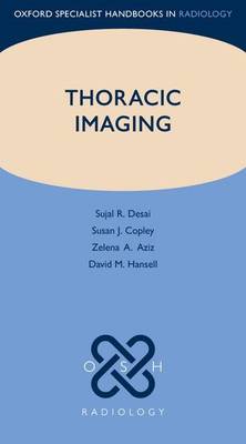
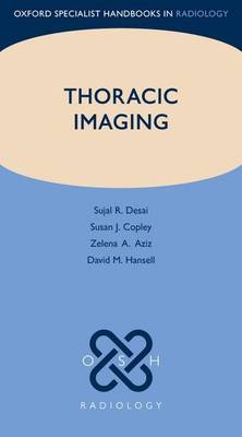
Imaging tests are integral to the management of patients with suspected lung disease. Staggering advances in radiological technology have been associated with an ever-increasing complexity, and so keeping up with terminology, understanding the significance of basic radiological signs, and appreciating the prognostic impact of findings on imaging tests, is not an easy undertaking for the physician. Until now, there has been a lack of practical, pocket-sized, but authoritative texts dealing principally with the imaging features of pulmonary disease. Covering the essential elements of pulmonary imaging in a concise and digestible format, Pulmonary Imaging deals with both the key principles of thoracic imaging, including a separate section on the common radiological terms used to describe pulmonary pathology, and the principal pathological compartments that are affected in specific disease processes. It is packed with over 600 high quality illustrations to highlight the important radiological features in the different diseases, with an emphasis on chest radiography (CXR) and computed tomography (CT) to mirror routine clinical practice.
Specificaties
Betrokkenen
- Auteur(s):
- Uitgeverij:
Inhoud
- Aantal bladzijden:
- 520
- Taal:
- Engels
- Reeks:
Eigenschappen
- Productcode (EAN):
- 9780199560479
- Verschijningsdatum:
- 5/12/2012
- Uitvoering:
- Paperback
- Formaat:
- Trade paperback (VS)
- Afmetingen:
- 99 mm x 173 mm
- Gewicht:
- 385 g

Alleen bij Standaard Boekhandel
+ 319 punten op je klantenkaart van Standaard Boekhandel
Beoordelingen
We publiceren alleen reviews die voldoen aan de voorwaarden voor reviews. Bekijk onze voorwaarden voor reviews.


