Je cadeautjes zeker op tijd in huis hebben voor de feestdagen? Kom langs in onze winkels en vind het perfecte geschenk!
- Afhalen na 1 uur in een winkel met voorraad
- Gratis thuislevering in België vanaf € 30
- Ruim aanbod met 7 miljoen producten
Je cadeautjes zeker op tijd in huis hebben voor de feestdagen? Kom langs in onze winkels en vind het perfecte geschenk!
- Afhalen na 1 uur in een winkel met voorraad
- Gratis thuislevering in België vanaf € 30
- Ruim aanbod met 7 miljoen producten
Zoeken
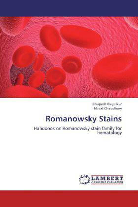
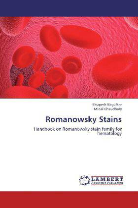
Romanowsky Stains
Handbook on Romanowsky stain family for hematology
Bhupesh Bagulkar, Minal Chaudhary
Paperback | Engels
€ 48,45
+ 96 punten
Omschrijving
In recent years, immunophenotying and cytogenetic analysis have become increasingly important in characterizing hematological neoplasms whilst the role of cytochemistry has diminished. Despite advances in recent years careful microscopical examination of romanowsky stained peripheral blood and bone marrow films remains fundamental in hematological diagnosis. Since the nineteenth century Romanowsky stains such as Giemsa's and Leishman's have provided the basis of recognizing, and indeed categorizing, the normal and pathological cells of peripheral blood and bone marrow. An advantage of Romanowsky staining for routine hematology is the highly polychrome image produced. Formation of the purple colour, i.e. the Romanowsky-Giemsa effect (RGE), is peculiar to these stains. So distinguishing between nuclear chromatin and basophilic cytoplasm, and between granules and cytoplasm, is harder with routine haematoxylin and eosin (H & E) than with Romanowsky staining.
Specificaties
Betrokkenen
- Auteur(s):
- Uitgeverij:
Inhoud
- Aantal bladzijden:
- 56
- Taal:
- Engels
Eigenschappen
- Productcode (EAN):
- 9783659265174
- Uitvoering:
- Paperback
- Afmetingen:
- 150 mm x 220 mm

Alleen bij Standaard Boekhandel
+ 96 punten op je klantenkaart van Standaard Boekhandel
Beoordelingen
We publiceren alleen reviews die voldoen aan de voorwaarden voor reviews. Bekijk onze voorwaarden voor reviews.


