- Afhalen na 1 uur in een winkel met voorraad
- Gratis thuislevering in België vanaf € 30
- Ruim aanbod met 7 miljoen producten
- Afhalen na 1 uur in een winkel met voorraad
- Gratis thuislevering in België vanaf € 30
- Ruim aanbod met 7 miljoen producten
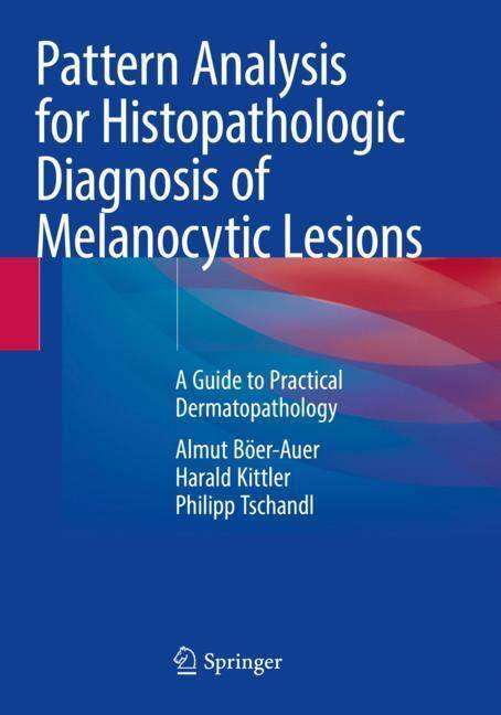
Pattern Analysis for Histopathologic Diagnosis of Melanocytic Lesions
A Guide to Practical Dermatopathology
Almut Böer-Auer, Harald Kittler, Philipp TschandlOmschrijving
Pattern analysis is a powerful method that changed dermatopathology, nowadays an indispensable tool in the diagnostic workup of inflammatory and neoplastic lesions. The diagnosis of melanocytic lesions can also be mastered by pattern analysis, which is the link between pathology, dermatoscopy, and clinical dermatology and supports the integration of all views. The histopathologic diagnosis of melanocytic lesions can be challenging for novices and experts alike. While classifications of melanocytic lesions come and go, pattern analysis is timeless; it can be assigned to any classification, current or future, and provides a framework that allows to address complex and uncertain cases in a repeatable manner. While uncertainty cannot be totally eliminated, pattern analysis helps to express this uncertainty in a meaningful way. Written by expert dermatopathologists with experience in dermatoscopy, this book is dedicated to young colleagues and to those who have not yet settled on one of the competing schools of thought; it is intended as a practical guide to help making correct observations, to describe them with a well-defined terminology, and to yield critical decisions in the face of incomplete or conflicting information. The illustrations contained in the volume are all original pictures in high-quality and full-color: reproductions of histopatological cuts in low and high magnification will assist pathologists, dermatologists, and dermatopathologists in interpreting histological slides of melanocytic skin lesions.
Specificaties
Betrokkenen
- Auteur(s):
- Uitgeverij:
Inhoud
- Aantal bladzijden:
- 272
- Taal:
- Engels
Eigenschappen
- Productcode (EAN):
- 9783031076688
- Verschijningsdatum:
- 17/12/2023
- Uitvoering:
- Paperback
- Formaat:
- Trade paperback (VS)
- Afmetingen:
- 178 mm x 254 mm
- Gewicht:
- 671 g

Alleen bij Standaard Boekhandel
Beoordelingen
We publiceren alleen reviews die voldoen aan de voorwaarden voor reviews. Bekijk onze voorwaarden voor reviews.


