
- Afhalen na 1 uur in een winkel met voorraad
- Gratis thuislevering in België vanaf € 30
- Ruim aanbod met 7 miljoen producten
- Afhalen na 1 uur in een winkel met voorraad
- Gratis thuislevering in België vanaf € 30
- Ruim aanbod met 7 miljoen producten


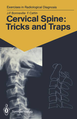
Cervical Spine: Tricks and Traps
60 Radiological Exercises for Students and Practitioners
Jean-Francois Bonneville, Francoise Cattin
€ 52,95
+ 105 punten
Omschrijving
The cervical spine is always examined initially using standard radiographs, which often provide a sufficient basis for diagno sis. Malformations, tumours, more frequently trauma, rheuma tism, and even plain neck pain require a radiological exam ination. The interpretation of radiological images is often difficult because of overlapping pieces of bone, the summation phe nomena and the diversity of projections. In this book, two- or three-dimensional CTscans accompany the standard radiographs, serving as an excellent aid for com prehension. It is almost as if the reader actually had the bones shown in the radiographs in his hands. From then on, everything becomes easy, superimpositions vanish, traps come to light, anatomy triumphs, and the images takes on life. Besanl$on, 1990 J .-F. BONNEVILLE, F. CATTIN v Contents Introduction 1 Iconography and Text with Corresponding Schemes 2 Subject Index . . . . . . . . . . . . . . . . . . .. . 123 VII Normal cervical spine: 3-D imaging 1 1 2 Case 1 1 Our starting point is a normal lateral radiograph of the cervical spine. It will serve as a guide and point of reference in our task of comprehending the interrelationships between the structures of the cervical spine. This radiograph correctly includes the entire area from the base of the skull to the cervicothora cic junction. The soft tissues are clearly visible anteriorly, as are the extremities of the spinous processes posteriorly. In this case, with the subject looking straight ahead, is a slight, regular, physiological lordosis ofthe cervical column."
Specificaties
Betrokkenen
- Auteur(s):
- Illustrator(s):
- Uitgeverij:
Inhoud
- Aantal bladzijden:
- 124
- Taal:
- Engels
- Reeks:
Eigenschappen
- Productcode (EAN):
- 9783540176831
- Verschijningsdatum:
- 4/05/1990
- Uitvoering:
- Paperback
- Formaat:
- Trade paperback (VS)
- Afmetingen:
- 133 mm x 203 mm
- Gewicht:
- 149 g


Alleen bij Standaard Boekhandel
+ 105 punten op je klantenkaart van Standaard Boekhandel
E-BOOK ACTIE


Beoordelingen
We publiceren alleen reviews die voldoen aan de voorwaarden voor reviews. Bekijk onze voorwaarden voor reviews.