- Afhalen na 1 uur in een winkel met voorraad
- Gratis thuislevering in België vanaf € 30
- Ruim aanbod met 7 miljoen producten
- Afhalen na 1 uur in een winkel met voorraad
- Gratis thuislevering in België vanaf € 30
- Ruim aanbod met 7 miljoen producten
Zoeken
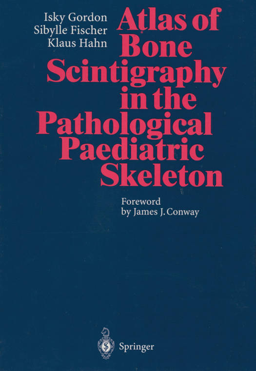
Atlas of Bone Scintigraphy in the Pathological Paediatric Skeleton
Under the Auspices of the Paediatric Committee of the European Association of Nuclear Medicine
Isky Gordon, Sibylle Fischer, Klaus Hahn
Paperback | Engels
€ 105,45
+ 210 punten
Omschrijving
This very practical "how-to" guide comprehensively covers both the common and less common pathologies affecting the paediatric skeleton. It provides clear explanations of the materials and instrumentation, as well as teaching points, technical comments, discussions, and the avoidance of pitfalls. The images presented here have been produced using whole-body scanning, gamma-camera, high-resolution spot images, pinhole and SPECT, as well as three-phase bone scans - each procedure backed by indications for its use. These 350 illustrations thus allow the paediatrician, orthopaedic surgeon, radiologist and nuclear medicine physician a comparison with their own images as well as with the "normal" images presented in the authors' companion volume, Atlas of Bone Scintigraphy in the Developing Paediatric Skeleton.
Specificaties
Betrokkenen
- Auteur(s):
- Uitgeverij:
Inhoud
- Aantal bladzijden:
- 343
- Taal:
- Engels
Eigenschappen
- Productcode (EAN):
- 9783642646751
- Verschijningsdatum:
- 16/11/2013
- Uitvoering:
- Paperback
- Formaat:
- Trade paperback (VS)
- Afmetingen:
- 216 mm x 279 mm
- Gewicht:
- 834 g
Alleen bij Standaard Boekhandel
+ 210 punten op je klantenkaart van Standaard Boekhandel
Beoordelingen
We publiceren alleen reviews die voldoen aan de voorwaarden voor reviews. Bekijk onze voorwaarden voor reviews.


